
Explore Virtual Dissection with Anatomage Tables at Simmons University
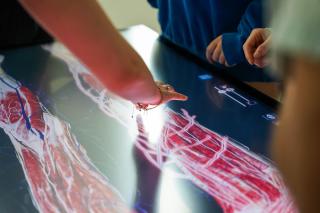
How Virtual Dissection Enhances Health Education
For decades, cadaver dissection has served as the foundational experience for students pursuing careers in medicine, physical therapy, and nursing. While physical specimens provide an irreplaceable realistic view of human anatomy, they come with significant limitations. Specimens are expensive to maintain, availability is limited, and once a structure is cut or removed, it cannot be easily restored for further study. Virtual dissection addresses these challenges by providing a sustainable, repeatable, and highly detailed alternative that complements traditional laboratory work.
As institutions across the country look to modernize their curricula, the integration of advanced visualization tools has become a prominent topic in USA news regarding higher education. These tools allow students to practice repeatedly without the risk of damaging a physical specimen. By bridging the gap between textbook diagrams and actual human bodies, virtual dissection platforms significantly improve a student’s ability to understand complex spatial relationships before they ever enter a clinical setting.
Explore our related articles for further reading on how technology is shaping modern degree programs.
Defining the Technology Behind Anatomage Tables
Anatomage Tables are life-size, interactive touchscreen systems designed specifically for studying human anatomy. Visually, the device resembles a traditional operating table, but instead of a physical body, it features a large, high-resolution display. The system uses advanced 3D visualization software to render fully segmented models derived directly from real human cadavers.
The technology was developed by a medical equipment manufacturer founded in 2004 and headquartered in Santa Clara, California. The company’s primary objective is to make science accessible and interactive for health science majors. Rather than relying on artistic renderings, the tables are built from high-resolution full-body CT and MRI scans of actual human donors. This approach ensures that the digital models preserve the natural anatomical variations found in real life, providing students with an authentic learning experience.
From Real Donors to Digital Models
Creating these highly detailed digital models is a rigorous process. Over more than two decades, the developers have digitally reconstructed over two million individual anatomical structures. The resulting software library contains more than 1,500 CT and MRI cases, each accompanied by clinical notes, 3D reconstructions, and examples of common diseases, traumatic injuries, and rare conditions.
Students and instructors can review the detailed medical histories of the original donors. This feature allows users to see exactly how specific diseases, such as cancer or cardiovascular issues, affected the donor’s body systems. By studying pathology in a 3D space, students gain a much deeper understanding of how anatomical structures respond to illness and injury.
Practical Applications in Anatomy and Physiology
Using an Anatomage Table, students can visualize muscles, nerves, blood vessels, ligaments, and organs in three dimensions. The interactive interface allows users to strip away layers of the body—skin, fat, muscle, and bone—individually or collectively. Users can rotate the body 360 degrees, zoom in on microscopic structures, and create cross-sectional views that mimic the appearance of actual CT scans.
One of the most valuable features for health education is the ability to animate joints. By moving a digital joint, students can observe exactly how muscles contract and relax during movement, and how ligaments and tendons provide stability. This dynamic view of anatomy is particularly beneficial for students studying physical therapy, sports medicine, orthopedics, and rehabilitation, where understanding biomechanics is just as important as knowing static anatomy.
Share your experiences in the comments below regarding how hands-on technology has impacted your study habits.
Simmons University Integrates Anatomage Tables into Nursing Education
Simmons University in Boston, Massachusetts, has actively adopted this technology to prepare students for demanding careers in healthcare. Beginning in the summer semester of 2026, students enrolled in the Simmons Doctor of Physical Therapy (DPT) degree program started using the tables alongside traditional cadaver dissection. Following this initial rollout, nursing education at Simmons is scheduled to integrate the technology during the 2026/2027 academic year.
The implementation of these tables directly impacts how faculty teach anatomy. In a traditional cadaver lab, a significant amount of time is spent simply locating and exposing structures before discussing their function. With the Anatomage Table, students can locate structures instantly. This efficiency allows instructors to dedicate more class time to discussing physiological function, clinical relevance, and pathological conditions, ultimately providing a richer educational experience.
Advantages for Physical Therapy Students
For Doctor of Physical Therapy students, understanding the musculoskeletal system requires more than memorizing bone landmarks. It requires an intimate knowledge of how force is generated across joints and how injuries compromise that function. The Anatomage Table allows DPT students to isolate specific muscle groups, trace nerve pathways from the spinal cord to the extremities, and visualize the exact mechanics of common injuries like rotator cuff tears or anterior cruciate ligament (ACL) ruptures. By analyzing these pathologies digitally, students can develop more effective, targeted treatment plans.
Schedule a free consultation to learn more about the DPT program requirements and clinical opportunities.
The Broader Impact on USA News and Health Education Trends
The adoption of virtual dissection technology at institutions like Simmons University reflects a broader trend in USA news concerning the future of STEM and medical education. As class sizes grow and the demand for highly trained healthcare professionals increases, universities must find ways to scale their anatomy education without compromising quality. Virtual dissection tables provide a scalable solution, allowing multiple small groups to study the same case simultaneously or review different pathologies side-by-side.
Furthermore, these systems offer an ethical and logistical advantage. While physical cadaver labs remain essential, the ability to study rare pathologies or congenital anomalies digitally—without needing to source specific physical specimens—ensures that all students receive a comprehensive education. This balance of traditional methods and cutting-edge technology is quickly becoming the gold standard for competitive health science programs.
Combining Technology with Traditional Clinical Labs
It is important to note that Anatomage Tables are designed to complement, rather than replace, hands-on clinical experience. At Simmons University, students do not rely on screens alone. They benefit from a comprehensive suite of learning environments designed to build practical skills.
For example, nursing students train in an 11,000-square-foot Nursing Simulation Lab. This controlled clinical setting features high-fidelity medical manikins, realistic hospital equipment, and simulated patient scenarios that build critical thinking and clinical judgment. Similarly, physical therapy students utilize dedicated Labs for Physical Therapy equipped with modern therapeutic equipment.
Simmons’ location in Boston further enhances these hands-on experiences. The university is situated just minutes away from the Longwood Medical Area, home to world-renowned institutions such as Brigham and Women’s Hospital, Beth Israel Deaconess Medical Center, Boston Children’s Hospital, and Dana-Farber Cancer Institute. This proximity provides students with unparalleled opportunities for clinical placements, internships, and observational experiences in some of the best healthcare facilities in the country.
Have questions? Write to us to find out how you can visit the simulation labs in person.
Evaluate Your Options for Advanced Health Degrees
Choosing the right university for your health science degree requires looking closely at the facilities and technologies available to undergraduate and graduate students. Simmons University offers a range of respected, women-centered undergraduate and co-educational graduate health degrees. Programs include Health and Exercise Science, a Bachelor of Science in Nursing (BSN), a Master of Science in Nursing (MSN) for BSN holders, Nutrition and Health Promotion, and the Doctor of Physical Therapy.
Integrating tools like the Anatomage Table into these curricula ensures that graduates are not only clinically competent but also technologically fluent. As healthcare continues to evolve, providers who are comfortable interpreting digital imaging, understanding 3D spatial anatomy, and utilizing interactive diagnostic tools will be well-positioned to lead in their respective fields.
Submit your application today to begin your journey in a technologically advanced health science program.